Kadrmas Eye Care New England
  • Doctors
    • Eddie Kadrmas, MD
    • Richard Strecker, MD
    • Daniel J. Townsend, MD
    • Aimee Gray, OD
    • Ashley Sousa, OD
  • Patients
    • CV-19 Statement
    • Eye Conditions >
      • Cataracts
      • Common Eye Problems >
        • Allergies
        • Amblyopia
        • Astigmatism
        • Blepharitis
        • Chalazion / Stye
        • Conjunctivitis
        • Corneal Abrasion
        • Floaters & Flashes
        • Hyperopia / Farsightedness
        • Myopia / Nearsightedness
        • Presbyopia
        • Strabismus
      • Corneal Diseases & Disorders >
        • Corneal Ulcers
        • Fuchs’ Dystrophy
        • Herpes Zoster / Shingles
        • Keratitis
        • Keratoconus
        • Pterygium
      • Diabetic Eye Disease
      • Dry Eye Syndrome
      • Eyelid Diseases & Disorders >
        • Dermatochalasis
        • Ectropion
        • Entropion
        • Eyelid Lesion
        • Ptosis
        • Tear Duct Obstruction
        • Tumor
      • Eye Trauma / Injury >
        • Blunt Trauma
        • Chemical Injury
        • Penetrating / Perforating Injury
      • Glaucoma >
        • Open Angle Glaucoma >
          • iStent
        • Narrow Angle Glaucoma
        • Neovascular Glaucoma
        • Inflammatory Glaucoma
      • Macular Diseases & Disorders >
        • Macular Degeneration
        • Macular Hole
        • Macular Pucker
      • Neurological Eye Diseases >
        • Blepharospasm
        • Double Vision
        • Ischemic Optic Neuropathy
        • Stroke / Visual Field Defect
        • Temporal Arteritis
      • Retinal Diseases & Disorders >
        • Retinal Tears & Detachments
        • Retinal Vascular Diseases
      • Uveitis & Iritis
    • Resources >
      • Eye Health & Care Blog
      • Retina Digest
    • Videos >
      • Eye Anatomy
      • Cataract
      • Laser Cataract Surgery
      • Children's Vision
      • Common Eye Problems
      • Corneal Conditions
      • Diabetic Eye Disease
      • Dry Eye Syndrome
      • Glaucoma
      • Laser & Refractive Eye Surgery
      • LASIK Over 45
      • LASIK Under 45
      • Macular Degeneration
      • Plastic Eye Surgery
      • Retinal Conditions
      • Refractive Errors
      • Routine Eye Exams
    • Forms & Policies >
      • New Patient Forms
      • HIPAA Policy
      • Referral Forms
    • Financial Information
  • Services
    • Routine Eye Exams >
      • Children
      • Adults
    • Hearing Therapy
    • Specialized Eye Exams >
      • Dilated Eye Exam / Slit Lamp Examination
      • Fluorescein Angiogram
      • Indocyanine Green Angiogram
      • Nerve Fiber Analysis
      • Optical Coherence Tomography
      • Refraction
      • Tonometry
      • Visual Acuity Testing
      • Visual Field Testing
    • Medical, Injection & Surgical / Laser Eye Care
    • Contact Lenses >
      • Contact Lens Types
      • Contact Lens Care & Safety
    • Refractive Eye Surgery >
      • LASIK Eye Surgery
      • PRK
      • Clear Lens Extraction
      • Implantable Collamer Lenses
    • Oculoplastic Surgery & Cosmetic Procedures >
      • Blepharoplasty
      • BOTOX®
      • Restylane®
  • Locations
    • Plymouth
    • Wareham
    • Mashpee
    • Surgery Centers
  • About
    • News
    • Affiliations
    • Patient Reviews
  • Careers
  • Contact
To Schedule Your Eye Appointment, Please Call 1-508-746-8600

Eye Conditions

Age-Related Macular Degeneration (AMD)

View Video
 

What Is Age-Related Macular Degeneration?

The macula, located in the center of the retina, is the portion of the eye responsible for your fine, detailed vision. The cells of the macula, known as photoreceptors, convert light into electrical images. The retina then sends these images via the optic nerve to the brain. A healthy macula allows you to do the daily activities you take for granted such as driving, reading, sewing and other detailed work.
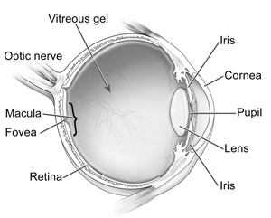
Eye Anatomy
Anatomy of the Eye
Photo Credit: National Eye Institute, National Institutes of Health

Age-related macular degeneration (AMD) is a disease associated with growing older that gradually destroys the macula. In some cases, the disease progresses very slowly and you may be unaware of the gradual deterioration of your central vision. In others, the deterioration can be rapid and can lead to loss of vision in both eyes.

People who suffer from AMD retain their peripheral vision. However, you may be unable to clearly see the object you are focusing on. In addition to the loss of central vision, you may also lose the ability to see color, or colors may appear to be less bright. You may find it increasingly difficult to recognize faces and your overall vision may seem hazy.

The Foundation Fighting Blindness notes that macular degeneration is the leading cause of blindness in people over the age of 55. In addition, the risk of developing macular degeneration increases as you age, most often affecting people in their 60s and 70s.

Who Is at Risk for Macular Degeneration?

As people age, everyone is at risk of developing macular degeneration.  Although AMD may occur during middle age, the National Eye Institute reports that people over the age of 60 are at greater risk than any other age group.
  
Normal Vision
Normal Vision
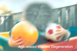
Age-related Macular Degeneration
Photo Credits: National Eye Institute, National Institutes of Health
If you are middle-aged, study results also show that you have about a 2-percent risk of getting AMD. However, your risk increases to nearly 30 percent if you are over the age of 75.

Other risk factors include smoking, obesity and family history. If you are white, you are much more likely to lose vision from AMD than if you are African American. In addition, women are at greater risk than men.

How is Macular Degeneration Detected?

Macular degeneration is diagnosed during a comprehensive eye examination, which will include:
  • Visual acuity test: Testing your vision at various distances
  • Dilated eye exam: Examining your retina and the back of your eye
  • Tonometry: Measuring your intraocular pressure
​
Your doctor may also use an Amsler Grid, a series of intersecting lines that resemble a checkerboard. A person with normal vision will see the lines as straight, while you would see the lines as wavy if you have AMD.

If your doctor suspects that you may have the wet type of macular degeneration, he may also suggest special tests, called fluorescein angiography, indocyanine green angiography or optical coherence tomography.

Once the testing is complete, the doctor will discuss the results with you. Since there are two types of AMD, dry AMD and wet AMD, specific treatment options will be reviewed with you.

Types of Age-Related Macular Degeneration

Select from the following links to learn more about the two types of age-related macular degeneration:
  • Dry Macular Degeneration
  • Wet Macular Degeneration

Treatment of Macular Degeneration

Select from the following options to learn more about treatment of macular degeneration: 
  • Medical Treatment for Macular Degeneration
  • Therapeutic Injections for Macular Degeneration
  • Laser Treatment for Macular Degeneration

Macular Degeneration Specialist at Kadrmas Eye Care New England

Meet our ophthalmologist who specializes in the treatment of macular degeneration:
  • Eddie F. Kadrmas, MD, PhD​

Back to Macular Diseases and Disorders Page

Kadrmas Eye Care New England

55 Commerce Way, Plymouth, MA 02360
14 Tobey Road, Wareham, MA 02571
133 Falmouth Road (Rt 28), Mashpee, MA 02649
Phone Number:
​1-508-746-8600

Hours:
Monday through Friday - 
8 AM - 4:30 PM 
© 2015-2021 Kadrmas Eye Care New England • All Rights Reserved
KadrmasEyeCare.com Privacy Policy • Disclaimer of Medical Liability
Proudly powered by Weebly
Design by DivTag Templates