Kadrmas Eye Care New England
  • Doctors
    • Eddie Kadrmas, MD
    • Richard Strecker, MD
    • Daniel J. Townsend, MD
    • David Lotufo, MD
    • Aimee Gray, OD
    • Ashley Sousa, OD
  • Patients
    • CV-19 Statement
    • Eye Conditions >
      • Cataracts
      • Common Eye Problems >
        • Allergies
        • Amblyopia
        • Astigmatism
        • Blepharitis
        • Chalazion / Stye
        • Conjunctivitis
        • Corneal Abrasion
        • Floaters & Flashes
        • Hyperopia / Farsightedness
        • Myopia / Nearsightedness
        • Presbyopia
        • Strabismus
      • Corneal Diseases & Disorders >
        • Corneal Ulcers
        • Fuchs’ Dystrophy
        • Herpes Zoster / Shingles
        • Keratitis
        • Keratoconus
        • Pterygium
      • Diabetic Eye Disease
      • Dry Eye Syndrome
      • Eyelid Diseases & Disorders >
        • Dermatochalasis
        • Ectropion
        • Entropion
        • Eyelid Lesion
        • Ptosis
        • Tear Duct Obstruction
        • Tumor
      • Eye Trauma / Injury >
        • Blunt Trauma
        • Chemical Injury
        • Penetrating / Perforating Injury
      • Glaucoma >
        • Open Angle Glaucoma >
          • iStent
        • Narrow Angle Glaucoma
        • Neovascular Glaucoma
        • Inflammatory Glaucoma
      • Macular Diseases & Disorders >
        • Macular Degeneration
        • Macular Hole
        • Macular Pucker
      • Neurological Eye Diseases >
        • Blepharospasm
        • Double Vision
        • Ischemic Optic Neuropathy
        • Stroke / Visual Field Defect
        • Temporal Arteritis
      • Retinal Diseases & Disorders >
        • Retinal Tears & Detachments
        • Retinal Vascular Diseases
      • Uveitis & Iritis
    • Resources >
      • Eye Health & Care Blog
      • Retina Digest
    • Videos >
      • Eye Anatomy
      • Cataract
      • Laser Cataract Surgery
      • Children's Vision
      • Common Eye Problems
      • Corneal Conditions
      • Diabetic Eye Disease
      • Dry Eye Syndrome
      • Glaucoma
      • Laser & Refractive Eye Surgery
      • LASIK Over 45
      • LASIK Under 45
      • Macular Degeneration
      • Plastic Eye Surgery
      • Retinal Conditions
      • Refractive Errors
      • Routine Eye Exams
    • Forms & Policies >
      • New Patient Forms
      • HIPAA Policy
      • Referral Forms
    • Financial Information
  • Services
    • Routine Eye Exams >
      • Children
      • Adults
    • Hearing Therapy
    • Specialized Eye Exams >
      • Dilated Eye Exam / Slit Lamp Examination
      • Fluorescein Angiogram
      • Indocyanine Green Angiogram
      • Nerve Fiber Analysis
      • Optical Coherence Tomography
      • Refraction
      • Tonometry
      • Visual Acuity Testing
      • Visual Field Testing
    • Medical, Injection & Surgical / Laser Eye Care
    • Contact Lenses >
      • Contact Lens Types
      • Contact Lens Care & Safety
    • Refractive Eye Surgery >
      • LASIK Eye Surgery
      • PRK
      • Clear Lens Extraction
      • Implantable Collamer Lenses
    • Oculoplastic Surgery & Cosmetic Procedures >
      • Blepharoplasty
      • BOTOX®
      • Restylane®
  • Locations
    • Plymouth
    • The Pinehills
    • East Wareham
    • Mashpee
    • Surgery Centers
  • About
    • News
    • Affiliations
    • Patient Reviews
  • Careers
  • Contact
To Schedule Your Eye Appointment, Please Call 1-508-746-8600

Eye Conditions

Retinal Tears and Detachments

View Video
 

What Are Retinal Tears and Detachments?

Retinal tears and detachments can result in blindness if not treated promptly. Therefore, if you have any of the symptoms listed below, you should contact our office immediately for an appointment. Retinal tears and detachments are considered to be ocular emergencies, and you should be evaluated by a doctor within 24 hours.

The eye is like a camera. The lens at the front of your eye focuses light onto your retina, the thin nerve layer at the back of the eye that transmits images via the optic nerve to your brain.
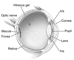
Anatomy of the Eye
Anatomy of the Eye
Photo Credit: National Eye Institute, National Institutes of Health

The vitreous gel is the fluid that fills our eye and is attached to the retina. This vitreous fluid helps the eye maintains its shape. When the vitreous gel changes shape as we get older, it tends to shrink and pull away from the retina. This shrinkage can cause the retina to tear or pull away from the back of the eye, usually in peripheral areas. 

Picture
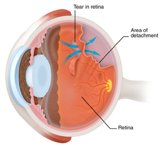
When this happens, fluid can leak through the retina and lift it off the back of the eye. Once the retina begins to separate from the back of the eye, it usually continues until most or all of it has detached. If the retina detaches, it is no longer sensitive to light and its light receptors will no longer be able to send images via the optic nerve to the brain. This results in loss of vision. Fortunately, prompt treatment can repair tears and reattach the retina.

Who Is at Risk for Retinal Tears and Detachments?

Although a retinal tear or detachment can occur at any age, the risk increases as we reach middle-age or older. This is due to normal shrinkage of the vitreous gel.

However, there are other factors that are associated with an increased risk, including:
  • Being nearsighted: Your eye tends to be longer and the retina thinner, making it more susceptible to a tear 
  • Sustaining an eye injury or head injury
  • Having eye surgery
  • Being diabetic
  • Having an eye disease that causes swelling or bleeding
  • Having a family or personal history of retinal detachment

What Are the Symptoms of  Retinal Tears and Detachments?

The warning signs of a detached retina including:
  • Flashing lights
  • A rapid increase of new floaters
  • A veil or curtain moving across your vision
  • A shadow in your peripheral vision; on the side, or from above or below

While these symptoms do not always mean that you have a detached retina, you should call your doctor immediately. If you have a tear or a detached retina, prompt treatment can prevent serious damage to your vision.

How Are Retinal Tears and Detachments Detected?

A tear or a retinal detachment is diagnosed during a dilated eye examination. Drops will be placed in your eyes to dilate your pupil and the doctor will examine your retina using an ophthalmoscope. This examination will determine the severity of the detachment, and whether the macula, the area of the retina responsible for central vision, is involved. These examinations take place at all five of our office locations.

Treatment of Retinal Tears and Detachments

Select from the following options to learn more about treatment of retinal tears and detachments:
  • Laser Treatment of Retinal Tears and Detachments
  • Surgical Treatment of Retinal Tears and Detachments

Retina Specialist at Kadrmas Eye Care New England

Meet our ophthalmologist who specializes in the treatment of retinal tears and detachments: 
  • Eddie F. Kadrmas, MD, PhD

Back to Retinal Diseases and Disorders Page

Kadrmas Eye Care New England

55 Commerce Way, Plymouth, MA 02360
The Pinehills, 1 Village Green North, Plymouth, MA 02360
3119 Cranberry Highway, East Wareham, MA 02538
133 Falmouth Road (Rt 28), Mashpee, MA 02649
Phone Number:
​1-508-746-8600

Hours:
Monday through Friday - 
8 AM - 4:30 PM 
Early Evening Hours Available - Please Call for Details
© 2015-2021 Kadrmas Eye Care New England • All Rights Reserved
KadrmasEyeCare.com Privacy Policy • Disclaimer of Medical Liability
Proudly powered by Weebly
Design by DivTag Templates