
Surgical Treatment for Retinal Tears and Detachments
|
Early detection and treatment of retinal tears may be able to prevent a retinal detachment. However, once your retina has detached, surgical intervention is required to re-attach the retina. According to the Mayo Clinic, approximately 90 percent of retinal detachments can successfully be treated, although in some cases, a second procedure may be required. While there is no guarantee that normal vision is restored, an untreated retinal detachment will almost certainly lead to total vision loss in your eye.
Pneumatic RetinopexyThe first type of surgical procedure for a detached retina is called pneumatic retinopexy. This procedure is normally used if the detachment is located in the top half of the retina.
|
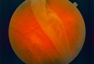
Detached Retina
|
Numbing drops will be applied to your eye. A small amount of aqueous fluid will be withdrawn from the eye. Your surgeon who is a retina specialist will then inject an expandable gas into the eye. Over the next several days, the gas bubble will expand and seal the retina tear. By holding the retina in place, the retina will reattach itself to the back of your eye. Laser treatment to permanently seal the tear will be performed 3-4 days later. Alternatively, cryotherapy may be applied to the tear at the time of the pneumatic retinopexy.
On the day of treatment, you will be welcomed by a staff member. The staff will help you prepare for your surgery by putting eye drops in your eye. You will be given a gown to wear over your street clothes during your actual surgery.
While your retina is healing, you may have to hold your head in a certain position for a few days to keep the bubble in place. Until the gas dissolves over a period of four to six (4-6) weeks, you will not be able to fly or be at high altitude. The air pressure of flying or being at high elevation can cause your eye pressure to rise rapidly and could result in serious complications. You should also avoid having general anesthesia during this period for another medical condition. Certain gases associated with general anesthesia can also cause similar problems.
While additional treatment may be required after pneumatic retinopexy, it can help you avoid more invasive surgical intervention.
After your treatment, you will rest in a comfortable reclining chair. Your family members may then join you in reviewing your follow-up care and schedule, including details on how to use the medicated eyedrops you may be given.
Although rare and treatable, complications of this treatment may include recurring retinal detachment, cataract formation, and glaucoma due to elevate eye pressure. For this reason, regular follow up visits will be required to monitor your progress.
Scleral Buckling
Scleral buckling is a common surgical treatment for a detached retina. This procedure is done under local anesthesia, and you will also be given a mild sedative. Your eye will be completely numbed, and you will remain comfortable during your surgical experience. This procedure is done at one of our Affiliated Ambulatory Surgery Centers (AASC).
You should not eat or drink anything after midnight the night before your procedure. On the day of treatment, you will be welcomed by a staff member. The staff will help you prepare for your surgery by putting eye drops in your eye. You will be given a gown to wear over your street clothes during your actual surgery.
Your surgeon will first treat the tear or detachment with cryopexy or laser. Depending on the severity of your detachment, he will then encircle all or part of the sclera, the white part of your eye, with a silicone band or buckle. This buckle will be stitched to the sclera and will hold the retina in place. Usually, the buckle will permanently remain in your eye.
Scleral buckling is normally successful in restoring vision. However, if the macula, the part of the retina responsible for central vision, is detached, the outcome can be uncertain. Many factors come into play including how long the retina and macula have been detached. For this reason, seeking treatment as soon as symptoms occur can be the key to preserving your vision.
In addition, the retina sometimes fails to reattach. This can be due to scar tissue that may develop behind or in front of the retina, both before and after the operation. If scar tissue does form, it can be removed in a surgical procedure called a vitrectomy. If the retina fails to attach for other reasons, pneumatic retinopexy may be used. This involves placing a gas bubble in your eye to help reattach the retina.
If your doctor inserts a gas bubble into your eye during this procedure, you may have to hold your head at an angled position for a few days to keep the bubble in place. Until the gas dissolves, usually over a period of four to six (4-6) weeks, you will not be able to fly or be at high altitude. The air pressure of flying or being at high elevation can cause your eye pressure to rise rapidly and could result in serious complications. You should also avoid having general anesthesia during this period for another medical condition. Certain gases associated with general anesthesia can also cause similar problems.
After your surgery, you will rest in a comfortable reclining chair. Your family members may then join you in reviewing your follow-up care and schedule. You may be given medication or eyedrops to prevent infection. Your surgeon will see you the next day after surgery to check how your eye is healing.
While complications are rare, they can include bleeding and glaucoma. For this reason, regular follow up visits will be required to monitor your progress.
Vitrectomy
Vitrectomy procedures may be the treatment of choice for many detachments. New technology has allowed the use of vitrectomy techniques to repair most detachments. If the eye has vitreous bleeding or if scar tissue has prevented the retina from successfully reattaching, your retina specialist will need to perform a vitrectomy to successfully reattach your retina.
This procedure is done under local anesthesia, and you will also be given a mild sedative. Your eye will be completely numbed, and you will remain comfortable during your surgical experience.
You should not eat or drink anything after midnight the night before your procedure. On the day of treatment, you will be welcomed by a staff member. The staff will help you prepare for your surgery by putting eye drops in your eye. You will be given a gown to wear over your street clothes during your actual surgery.
Your surgeon will look into your eye with a microscope. Tiny incisions will be made in the sclera, the white part of your eye. Microsurgical instruments are then used to perform various maneuvers.
Depending on the nature of your situation, your surgeon will do one or more of the following to ensure the best possible vision outcome:
- Remove cloudy or bloody vitreous
- Remove any scar tissue and return the retina to its normal position
- Remove any foreign object in your eye
- Treat the eye with a laser to seal the retinal tears and holes
- Use a heavy liquid to help reattach the retina
- Use pneumatic retinopexy to insert a gas bubble to hold your retina in place
- Utilize a scleral buckle to hold the retina in place
If your doctor inserts a gas bubble into your eye during this procedure, you will be instructed to keep your head in a certain position for a few weeks to keep the bubble in place. Until the gas dissolves, usually over a period of four to six (4-6) weeks, you will not be able to fly or be at high altitude. The air pressure of flying or being at high elevation can cause your eye pressure to rise rapidly and could result in serious complications. You should also avoid having general anesthesia during this period for another medical condition. Certain gases associated with general anesthesia can also cause similar problems.
After your surgery, you will rest in a comfortable reclining chair. Your family members may then join you in reviewing your follow-up care and schedule. You may be given medication or eyedrops to prevent infection. Your surgeon will see you the next day after surgery to check how your eye is healing.
Some discomfort is to be expected following surgery. You will wear an eye patch for a short time afterwards. It will take a month for the surface of your eye to fully heal. Depending on the status of your retina, it may take six months or more to attain your final visual result. You may need to avoid strenuous activity while the gas bubble is in place. It will be necessary for you to see your doctor at regular intervals during this time so that he can monitor your healing.
Any surgical procedure involves some risks. These risks include infection, bleeding, retinal re-detachment, high intraocular pressure and accelerated cataract formation. However, most of these conditions can be successfully treated. The potential risks associated with a vitrectomy are less than the expected benefits of preserving your vision.
An untreated detached retina can lead to loss of vision in your eye. Therefore, symptoms such as flashing lights, a rapid increase of new floaters, a veil or curtain moving across your vision or a shadow in your sideways or peripheral vision, symptoms that can be indications of a detached retina, should not be ignored. While they do not always mean that you have a detached retina, you should call your doctor immediately. If you do have a tear or a detachment, prompt treatment can prevent serious damage to your vision.