Kadrmas Eye Care New England
  • Doctors
    • Eddie Kadrmas, MD
    • Richard Strecker, MD
    • Daniel J. Townsend, MD
    • Aimee Gray, OD
    • Ashley Sousa, OD
  • Patients
    • CV-19 Statement
    • Eye Conditions >
      • Cataracts
      • Common Eye Problems >
        • Allergies
        • Amblyopia
        • Astigmatism
        • Blepharitis
        • Chalazion / Stye
        • Conjunctivitis
        • Corneal Abrasion
        • Floaters & Flashes
        • Hyperopia / Farsightedness
        • Myopia / Nearsightedness
        • Presbyopia
        • Strabismus
      • Corneal Diseases & Disorders >
        • Corneal Ulcers
        • Fuchs’ Dystrophy
        • Herpes Zoster / Shingles
        • Keratitis
        • Keratoconus
        • Pterygium
      • Diabetic Eye Disease
      • Dry Eye Syndrome
      • Eyelid Diseases & Disorders >
        • Dermatochalasis
        • Ectropion
        • Entropion
        • Eyelid Lesion
        • Ptosis
        • Tear Duct Obstruction
        • Tumor
      • Eye Trauma / Injury >
        • Blunt Trauma
        • Chemical Injury
        • Penetrating / Perforating Injury
      • Glaucoma >
        • Open Angle Glaucoma >
          • iStent
        • Narrow Angle Glaucoma
        • Neovascular Glaucoma
        • Inflammatory Glaucoma
      • Macular Diseases & Disorders >
        • Macular Degeneration
        • Macular Hole
        • Macular Pucker
      • Neurological Eye Diseases >
        • Blepharospasm
        • Double Vision
        • Ischemic Optic Neuropathy
        • Stroke / Visual Field Defect
        • Temporal Arteritis
      • Retinal Diseases & Disorders >
        • Retinal Tears & Detachments
        • Retinal Vascular Diseases
      • Uveitis & Iritis
    • Resources >
      • Eye Health & Care Blog
      • Retina Digest
    • Videos >
      • Eye Anatomy
      • Cataract
      • Laser Cataract Surgery
      • Children's Vision
      • Common Eye Problems
      • Corneal Conditions
      • Diabetic Eye Disease
      • Dry Eye Syndrome
      • Glaucoma
      • Laser & Refractive Eye Surgery
      • LASIK Over 45
      • LASIK Under 45
      • Macular Degeneration
      • Plastic Eye Surgery
      • Retinal Conditions
      • Refractive Errors
      • Routine Eye Exams
    • Forms & Policies >
      • New Patient Forms
      • HIPAA Policy
      • Referral Forms
    • Financial Information
  • Services
    • Routine Eye Exams >
      • Children
      • Adults
    • Hearing Therapy
    • Specialized Eye Exams >
      • Dilated Eye Exam / Slit Lamp Examination
      • Fluorescein Angiogram
      • Indocyanine Green Angiogram
      • Nerve Fiber Analysis
      • Optical Coherence Tomography
      • Refraction
      • Tonometry
      • Visual Acuity Testing
      • Visual Field Testing
    • Medical, Injection & Surgical / Laser Eye Care
    • Contact Lenses >
      • Contact Lens Types
      • Contact Lens Care & Safety
    • Refractive Eye Surgery >
      • LASIK Eye Surgery
      • PRK
      • Clear Lens Extraction
      • Implantable Collamer Lenses
    • Oculoplastic Surgery & Cosmetic Procedures >
      • Blepharoplasty
      • BOTOX®
      • Restylane®
  • Locations
    • Plymouth
    • Wareham
    • Mashpee
    • Surgery Centers
  • About
    • News
    • Affiliations
    • Patient Reviews
  • Careers
  • Contact
To Schedule Your Eye Appointment, Please Call 1-508-746-8600

Patient Resources - Eye Health & Care Blog

Common Causes of Vision Loss in Younger Adults: National Save Your Vision Month

3/9/2016

 
March is National Save Your Vision Month, and in our five-part blog series, we’re taking a look at common causes of vision loss in adulthood, eye exams, general eye health, and signs telling you to have your eyes checked.
​
In this second blog post, we explore common causes of vision loss in younger adults, both of which affect the retina:
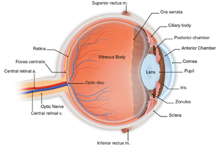
  1. Retinal Tears & Detachments
  2. Diabetic Retinopathy 
​​
Picture

Retinal Tears & Detachments

Picture
Retinal tears and detachments are optical emergencies that can cause blindness if not evaluated and treated promptly (within 24 hours). If you or a loved one experience any of the symptoms listed below, contact our office or your eye doctor immediately.
 
Retinal tears and detachments happen as the vitreous gel, the fluid fills the eye and helps it maintain its shape, changes shape as we get older. It tends to shrink and pull away from the retina, the thin nerve layer at the back of the eye at that accepts light and transmits images to the brain via the optic nerve. In some cases, this shrinkage of the vitreous gel can cause the retina to tear or pull away from the back of the eye, usually in the peripheral (side) areas.
 
When the retina tears or pulls away, fluid can leak through the retina and cause it to actually lift off the back of the eye. Once this process begins, it usually continues until most of or the entire retina has detached. If this happens, the light receptors in the retina will no longer be able to accept light or send images to the brain, which results in loss of vision. Fortunately, prompt treatment can repair tears in the retina and reattach the retina to the back of the eye, which is why it is so important to contact your eye doctor immediately if you or a loved one begin to experience any of the following symptoms or warning signs:
  • Flashing lights
  • A rapid increase of new floaters
  • A veil or curtain moving across your vision
  • A shadow in your peripheral vision, to the side or above or below
 
While these symptoms are warning signs, they do not always signify a retinal tear or detachment. However, due to the need to be evaluated and treated by an ophthalmologist who specializes in retinal tears and detachments promptly, don’t wait to call your eye doctor if you or a loved one develop any of these symptoms.
 
Certain people, such as people who are middle age and older, are at a higher risk for retinal tears and detachments due to normal shrinkage of the vitreous gel. In addition, the following increase risk of developing this serious condition:
  • Nearsightedness, as the eye tends to be longer and the retina thinner, making it more susceptible to a tear
  • An eye or head injury
  • Eye surgery
  • Diabetes
  • Any eye disease that causes edema (swelling) or bleeding in the eye
  • Family or personal history of retinal detachment
 
Your ophthalmologist can detect a retinal tear or detachment through a dilated eye examination, which allows the doctor to examine your retina using a device called an ophthalmoscope. This examination will help your eye doctor understand the severity of your condition and whether the macula, the area of the retina responsible for fine, detailed vision used for activities such as reading and writing, is involved. Further, it will help your eye surgeon understand whether laser or surgical treatment is right for your particular retinal tear or detachment.
 
To learn more about retinal tears and detachments, as well as treatment options for these optical emergencies, visit our retinal tears and detachment webpages. 

Diabetic Retinopathy

Picture
Diabetic retinopathy, caused by vascular changes in the retina (the light sensitive area in the back of the eye) related to diabetes, is one of the leading causes of vision loss and blindness in adults. In some cases, blood vessels in eye may swell and leak, whereas in other cases, abnormal new blood vessels may grow on the retina.
 
Diabetic retinopathy occurs in four stages:

  1. Stage 1 – Mild Nonproliferative Retinopathy: Non-painful swelling of the blood vessels of the retina like a small balloon (called a microaneurysm).
  2. Stage 2 – Moderate Nonproliferative Retinopathy: Some blood vessels that nourish the retina become blocked.
  3. Stage 3 – Severe Nonproliferative Retinopathy: Many more blood vessels supplying the retina become blocked, depriving the retina of much needed nourishment and signaling the retina to create new blood vessels in a process called neovascularization. Because these new vessels are weak, they may leak, and swelling or edema in the macula (the part of the eye that converts light into electrical signals) may occur.
  4. Stage 4 – Proliferative Retinopathy: Even more blood vessels are blocked and neovascularization increases. The new weak vessels grow along the retina and into the vitreous gel inside the eye, and their fragility can cause them to start to bleed into the center of the eye. Macular edema (swelling) is more likely to occur as well.
 
Vision loss can begin to occur at any stage of diabetic retinopathy. However, as the disease progresses and the macula is affected and bleeding worsens, many will notice vision changes in Stage 3 and blurry vision in Stage 4 diabetic retinopathy. If macular edema is present, fine vision used for reading, writing, and other detailed work may become diminished or lost. 
 
In addition to vision loss, black spots or flashes of light in central vision may become noticeable as diabetic retinopathy progresses. These can be symptoms of a detached retina or vitreous hemorrhage, both of which can cause a rapid loss of vision and constitute a medical emergency. If you notice any of these symptoms, you should see your ophthalmologist or eye doctor immediately. While it is not possible to restore lost vision, prompt treatment by your ophthalmologist who specializes in diabetic retinopathy can prevent further vision loss.
 
Everyone with diabetes is at risk for diabetic retinopathy, regardless of the type. However, people who have had diabetes for longer are at greater risk. If you have diabetes, you should have a dilated eye exam once a year. Because proliferative retinopathy and macular edema can occur without noticeable vision loss, your vision may be at risk without regular examination, and early detection and treatment of diabetic retinopathy can prevent vision loss.
 
If you are pregnant and have diabetes, you should see your eye doctor as soon as possible, as more frequent eye examinations to check for signs of diabetic retinopathy may be needed. If you have been diagnosed with diabetic retinopathy, you may also need dilated eye exams more frequently.
 
In addition to dilated eye exams, your eye doctor will perform a comprehensive eye examination to diagnose and monitor diabetic retinopathy, including:
  • Visual acuity test
  • Tonometry
  • Fluorescein angiogram
  • Optical coherence tomography
 
These tests will help your ophthalmologist understand your condition and determine the best course of treatment for you.
 
To learn more about diabetic retinopathy and medical, laser, therapeutic injection, and surgical treatments for this condition, please visit our diabetic eye disease webpages. 
​​

Specialists in Retinal Tears, Retinal Detachments & Diabetic Retinopathy

If you or a loved one is suffering from or has symptoms consistent with either of these conditions described above, please contact us. Our ophthalmologists, who specialize in these conditions as listed below, are here to help save your vision:
  • For Retinal Tears & Detachments: Eddie F. Kadrmas, MD, PhD
  • For Diabetic Retinopathy: Eddie F. Kadrmas, MD, PhD & Lawrence I. Rand, MD
​
Contact Us to Schedule an Appointment

Comments are closed.

    EYE HEALTH BLOG


    CLICK HERE to sign up for our eye health / care newsletter and have it delivered automatically to your inbox each month!

    Categories

    All
    AMD
    Astigmatism
    Cataract
    Children's Eye Health
    Color Blindness
    Conjunctivitis
    Contact Lens
    Cornea
    Cosmetic Eye Surgery
    Covid 19
    Diabetic Eye Disease
    Driving
    Dry Eyes
    Eye Allergies
    Eye Cancer
    Eye Exam
    Eye Health & Safety
    Eye Infections
    Eye Strain
    Glaucoma
    Injuries
    Low Vision
    Lupus
    Macula
    Myasthenia Gravis
    Organ Donation
    Retina
    Retinopathy
    Scleritis
    Senior Eye Health
    Sports
    Stroke
    Stye
    Uvea
    Vision Correction
    Vision Loss
    Women's Eye Health

    RSS Feed


    Picture

Kadrmas Eye Care New England

55 Commerce Way, Plymouth, MA 02360
14 Tobey Road, Wareham, MA 02571
133 Falmouth Road (Rt 28), Mashpee, MA 02649
Phone Number:
​1-508-746-8600

Hours:
Monday through Friday - 
8 AM - 4:30 PM 
© 2015-2021 Kadrmas Eye Care New England • All Rights Reserved
KadrmasEyeCare.com Privacy Policy • Disclaimer of Medical Liability
Proudly powered by Weebly
Design by DivTag Templates