|
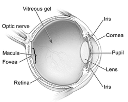
March is National Save Your Vision Month, and in our five-part blog series, we’re taking a look at common causes of vision loss in adulthood, eye exams, general eye health, and signs telling you to have your eyes checked. We’ll look at each of these conditions, including their causes and symptoms, in detail in this post. Age-Related Macular Degeneration (AMDAMD is an eye disease that is associated with age and that gradually destroys the macula, located in a part of the back of the eye called the retina. The macula is responsible for our fine, detailed vision that we use everyday for activities such as reading, writing, driving, knitting, sewing, and other detailed work. Because the macula is in the center of the retina, macular degeneration affects central vision. Although peripheral vision (side-vision) is retained, meaning someone with AMD will not go completely blind from macular degeneration, it may be difficult or impossible to clearly focus on or see an object, a person’s face, or words on a page. Further, ability to see color may be affected and overall vision may seem hazy. In some cases, the onset of AMD may be slow and lead to gradual deterioration of vision. In other cases, onset can be fast, and deterioration of vision can be rapid and lead to loss of vision in both eyes. As the name and the subject of this blog post indicate, aging is a risk factor for aged-related macular degeneration. While AMD can occur during middle age, people over 60 are at the greatest risk for developing this disease, and people over the age of 75 have a 30-percent chance of developing AMD. Additional risk factors for AMD include smoking, obesity, family history, Caucasian decent, and being female. There are two types of macular degeneration, dry macular degeneration and wet macular degeneration, which are diagnosed during a comprehensive eye examination. In addition, your eye doctor may use an Amsler Grid to assess your vision, as well as recommend special tests including fluorescein angiography, indocyanine green angiography, or optical coherence tomography. The results of these tests will help your ophthalmologist understand your particular condition, as well as appropriate treatments for your particular type of AMD. Because AMD can lead to loss of vision, it is important to understand these symptoms and contact your ophthalmologist immediately if any symptoms change or worsen. It is also important to have your eyes examined by your ophthalmologist regularly to understand the changes in your eyes and the effectiveness of treatments. To learn more about AMD and AMD treatment, please visit our AMD webpages, as well as our National AMD & Low Vision Awareness Month blog post series. In addition to detailed information, you will find videos that will help you better understand this condition. Cataracts A cataract, or a cloudy lens of the eye, is a common eye condition normally associated with aging. As we age, proteins in the lens of our eye begin to form into small clumps that can affect vision. The lens, located directly behind the iris and pupil, focuses light and images on the retina and allows vision to shift or adjust vision from up close to far away and vice versa. When affected by cataracts, the cloudy lens can cause difficulty with vision. A cataract reduces the sharpness of vision, particularly at night and with driving in the dark. Lights may seem too bright, or you may feel an uncomfortable glare or see halos around the light. These symptoms may also occur during the day in bright sunlight. In addition to cloudiness, the lens can become yellowish or brownish with a cataract, which can ability to see color. Colors may appear faded, and in more advanced or pronounced cases, distinguishing blues and purples may become difficult. A cataract may develop during the aging process, from eye trauma, as a result of surgery for another eye condition such as glaucoma, as a consequence of a systemic disease such as diabetes, or following radiation exposure. A cataract can develop in people as young as 40 or 50, but for most people, vision loss doesn’t begin to occur until after age 60. Diabetes, smoking, and drinking alcohol are linked to increased risk of developing cataracts. In addition, prolonged exposure to sunlight also increases risk. Sunglasses worn anytime out in the sun can help minimize this risk. A cataract or cataracts can be detected during a comprehensive eye examination, which includes a visual acuity test, dilated eye exam, and tonometry. Cataracts are treated with laser surgery, one of the most frequently performed surgical procedures in the U.S. and one of the safest and most successful. Your ophthalmologist who specializes in cataract surgery can speak with you about the procedure and help you find the right time to get your vision back. To learn more about cataracts and cataract surgery, please visit our cataract webpages, where you will find detailed information and educational videos. Glaucoma Glaucoma, a group of diseases that cause damage to the optic nerve (a bundle of nerve fibers at the back of the eye that connects and transfers images from the eye to the brain) is the second leading cause of blindness in adults in the U.S. Because glaucoma can cause loss of vision and blindness, it is important to know that it can be detected through regular eye examinations. Regular eye examinations are so important because there may be no obvious symptoms in the early stages of glaucoma, even though damage to the optic nerve is occurring. Eventually, as damage to the optic nerve progresses, blind spots will develop. By the time you become aware of these blind spots, the optic nerve has been significantly damaged. The good news is that early detection and treatment by your ophthalmologist is the key to preventing nerve damage and blindness. The Glaucoma Research Foundation recommends the following schedule for routine eye examinations:
For people at increased risk of glaucoma, the Glaucoma Research Foundation recommends annual testing starting between the ages of 35 and 40. While anyone can develop glaucoma at any age, a number of risk factors exist, including:
Glaucoma is diagnosed through a routine eye examination, including tonometry, gonioscopy, ophthalmoscopy, visual field testing, and in some cases, optical coherence tomography. These examinations will help your ophthalmologist determine the type of glaucoma (open angle, narrow angle, neovascular, or inflammatory) you have and the best treatment for your particular condition. Further, these eye examinations will need to be repeated at regular intervals to monitor changes in your condition. To learn more about glaucoma, visit our glaucoma webpage. To learn about the different types of glaucoma and their treatments, click the links below: Diabetic Retinopathy Diabetic retinopathy, caused by vascular changes in the retina (the light sensitive area in the back of the eye) related to diabetes, is one of the leading causes of vision loss and blindness in adults. In some cases, blood vessels in eye may swell and leak, whereas in other cases, abnormal new blood vessels may grow on the retina. Diabetic retinopathy occurs in four stages:
Vision loss can begin to occur at any stage of diabetic retinopathy. However, as the disease progresses and the macula is affected and bleeding worsens, many will notice vision changes in Stage 3 and blurry vision in Stage 4 diabetic retinopathy. If macular edema is present, fine vision used for reading, writing, and other detailed work may become diminished or lost. In addition to vision loss, black spots or flashes of light in central vision may become noticeable as diabetic retinopathy progresses. These can be symptoms of a detached retina or vitreous hemorrhage, both of which can cause a rapid loss of vision and constitute a medical emergency. If you notice any of these symptoms, you should see your ophthalmologist or eye doctor immediately. While it is not possible to restore lost vision, prompt treatment by your ophthalmologist who specializes in diabetic retinopathy can prevent further vision loss. Everyone with diabetes is at risk for diabetic retinopathy, regardless of the type. However, people who have had diabetes for longer are at greater risk. If you have diabetes, you should have a dilated eye exam once a year. Because proliferative retinopathy and macular edema can occur without noticeable vision loss, your vision may be at risk without regular examination, and early detection and treatment of diabetic retinopathy can prevent vision loss. If you are pregnant and have diabetes, you should see your eye doctor as soon as possible, as more frequent eye examinations to check for signs of diabetic retinopathy may be needed. If you have been diagnosed with diabetic retinopathy, you may also need dilated eye exams more frequently. In addition to dilated eye exams, your eye doctor will perform a comprehensive eye examination to diagnose and monitor diabetic retinopathy, including: These tests will help your ophthalmologist understand your condition and determine the best course of treatment for you. To learn more about diabetic retinopathy and medical, laser, therapeutic injection, and surgical treatments for this condition, please visit our diabetic eye disease webpages. Specialists in AMD, Cataracts, Glaucoma & Diabetic RetinopathyIf you or a loved one is suffering from or has symptoms consistent with any of these conditions described above, please contact us. Our ophthalmologists, who specialize in these conditions as listed below, are here to help save your vision:
Comments are closed.
|
EYE HEALTH BLOGCategories
All
Archives
April 2024
|
|
Kadrmas Eye Care New England
55 Commerce Way, Plymouth, MA 02360
14 Tobey Road, Wareham, MA 02571 133 Falmouth Road (Rt 28), Mashpee, MA 02649 |
Phone Number:
1-508-746-8600 Hours: Monday through Friday — 8 AM – 4:30 PM |


 RSS Feed
