
|
March is National Save Your Vision Month, and in our five-part blog series, we’re taking a look at common causes of vision loss in adulthood, eye exams, general eye health, and signs telling you to have your eyes checked.
While people with healthy eyes can have complete eye exams at regular intervals based on age, people at risk for certain eye conditions should have routine eye exams more frequently starting earlier in life. In particular, we want to look for and detect conditions that threaten vision such as diabetic retinopathy, glaucoma, cataracts, and macular degeneration as early as possible.
For people who have been diagnosed and are living with diabetes, we recommend having a yearly eye examination. This recommendation is regardless of age due to the risk of developing diabetic retinopathy. Whether you are 20 and have Type 1 diabetes or are 55 and have Type 2 diabetes, you should have your eyes checked annually. As people at risk of glaucoma, as well as other eye conditions such as cataracts and macular degeneration, have a better chance of preserving vision if they are detected early and monitored regularly by an ophthalmologist, we recommend understanding your risk, discussing it with your eye doctor, and determining a routine eye examination schedule that is right for you. People at greater risk for eye disease can have their eye health and vision assessed and monitored by a complete eye examination, which can be performed by a general ophthalmologist or optometrist. If any portion of the complete eye examination detects a potentially more serious or vision threatening eye condition, your eye doctor may recommend further examination and one or more of the following advanced / specialized studies for your eyes:
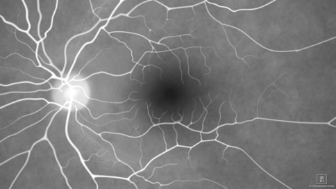
Fluorescein Angiogram (FA)
FA is a special test that helps your ophthalmologist diagnose many retinal conditions. Your ophthalmologist will inject dye into your eye arm, which will circulate through your body and into the blood vessels in your eyes. He or she will then use a special fundus camera (non x-ray technology) to take a series of pictures of the blood vessels your eyes over the period of approximately 10 minutes.
To learn more about FA, visit our FA webpage. Inodcyanine Green Angiogram (ICG)
Similar to FA, ICG is another special test that helps your ophthalmologist diagnose certain retinal conditions. However, this particular type of angiography is able to identify eye disease in the choroidal vessels behind the retina that may not be visible on FA. This test uses a different type of dye that is detected by a special camera that takes infrared images and takes about 20 minutes.
To learn more about ICG, visit our ICG webpage. Nerve Fiber Analysis
A nerve fiber analysis test is preformed by your ophthalmologist to detect any damage to the nerve fiber layer around the optic nerve caused by glaucoma. A painless scanning laser will measure the thickness of the nerve fiber layer, which is compared to a ‘normal’ profile of someone your age, as well as any results from previous nerve fiber analysis tests you’ve had in the past.
To learn more about this test, visit our nerve fiber analysis webpage. Optical Coherence Tomography (OCT)
OCT is a non-invasive examination that is used to visualize the 10 layers that make up the retina. It allows your ophthalmologist to assess the thickness of each layer to be measured and confirm a specific retinal diagnosis such as a macular hole, as well as to track progressive retinal diseases such as age-related macular degeneration (AMD). Further, OCT can measure the surface contour of the optic nerve and the thickness of the surrounding nerve fiber layer. These results are compared to a ‘normal’ profile of someone your age, any results from previous tests as in nerve fiber analysis. The results of one eye are compared to that of the opposite eye, as asymmetry between eyes can be a sign of early glaucoma as changes to the optic disc (where retinal nerve fibers converge and exit the back of the eye) or nerve fiber layer begin.
To learn more about OCT, visit our OCT webpage. Visual Field Testing
Through visual field testing, your ophthalmologist assesses your central and peripheral (side) vision. With one eye covered, you look into a small, semi-circular bowl and focus on a small light. Small dim lights are then projected on the center and sides of the bowl, and you will be asked to press a buzzer when you see these lights. Each eye will be tested separately, and both of your results are compared to a ‘normal’ profile of someone your age, as ell as any previous visual field tests you’ve had.
To learn more about this examination, visit our visual field testing page. Ophthalmologists Who Perform Specialized Eye Tests
If you are just establishing care with our practice, one of our general ophthalmologists or optometrists will typically preform your complete eye exam and discuss your risk factors and any symptoms with you. Based on the results of your complete eye exam, the eye doctor may recommend further tests to diagnosis and assess your eye condition, as well as refer you to one of our ophthalmologists who specializes in your particular condition. Complete eye exams are performed in all of our five locations on the South Shore and Cape Cod and can be scheduled by calling 508-746-8600.
To learn more about our specialized ophthalmologists, general ophthalmologists, and optometrists, visit their profile pages:
Comments are closed.
|
EYE HEALTH BLOGCategories
All
Archives
July 2024

|
 RSS Feed
|
Kadrmas Eye Care New England
55 Commerce Way, Plymouth, MA 02360
14 Tobey Road, Wareham, MA 02571 133 Falmouth Road (Rt 28), Mashpee, MA 02649 |
Phone Number:
1-508-746-8600 Hours: Monday through Friday — 8 AM – 4:30 PM |